Indian Journal of Physiology
and Pharmacology
and Pharmacology
Indian Journal of Physiology and Pharmacology |
 (252x320).jpg) |
Volume 58 - Number 2 April - 2014 (Current issue) ISSN 0019-5499 |
Effects of Age and Body Mass Index on Peak Expiratory Flow Rate
|
Peak expiratory flow rate (PEFR) is a measure of ventilatory capacity measured by peak flow meter. It is regarded as a basic physiological parameter for the diagnosis, follow up and treatment of patients with respiratory illnesses such as asthma, chronic bronchitis, and emphysema. This study establishes the relationship of PEFR with age and BMI in healthy adult males (N=300) of Kumaon region of Uttarakhand. Overall, the mean PEFR is 478.37+68.14. The age is significantly affecting the PEFR unlike BMI. Age as an independent predictor predicts the 72.3% variability (R is 72.3%)in PEFR while BMI > 23 as an independent predictor predicts only 1.4% variability (R2 is 0.014) .PEFR declines with advancing age due to degenerative changes in musculoskeletal system leading to decrease in respiratory muscle strength. PEFR shows some decline with high BMI in elderly age group. |
Pulmonary functions are generally determined by respiratory muscle strength, compliance of the thoracic cavity, airway resistance and elastic recoil of the lungs (1). Narrowing of airways is expressed in terms of various expiratory flow rates. Peak expiratory flow rate (PEFR) is one such parameter that can be easily measured by a peak flow meter and is a conventional tool for measure lung functions in field study (2). It is a fairly good indicator of bronchial hyper-responsiveness and does not require body-temperature-pressuresaturation correction (BTPS) (3). The primary aim of this study is to establish the effect of age and BMI on PEFR on Indian adult males who live in Kumaon region of Uttarakhand which comes in hilly region and considered as relatively pollution free in comparison to other places of India. |
The present study was undertaken in the department of Physiology at Government Medical College, Haldwani, Nainital. Three hundred healthy, nonsmoker males in the age range of 18-60 years volunteered for this study. Study area : Kumaon region of Uttarakhand. Study subjects : Residents of different villages of Kumaon region of Uttarakhand and area near the government medical college, Haldwani. Inclusion Criteria for Selection of study subjects :
Study period : November 2012 to February 2013. The peak expiratory flow rate was determined using Wright's Peak flow meter. The subjects were asked to stand in upright position with the peak flow meter held horizontally in front of their mouth and take a deep breath in and close the lips firmly around the mouthpiece, making sure that no air leaks around the lips. The subjects were asked to breathe out as hard and as fast as possible and the number indicated by the cursor was noted. The above sequence was repeated thrice in each patient. The highest of the three measurements was taken in to consideration for the analysis. The BMI was calculated using the formula- body weight (in kg)/height2 (in meter). Statistical analysis Statistical analysis was done using SPSS version 18 and MS Office excel 2007. Variables analysed were age, BMI and PEFR. Student's t test and Linear regression analysis was done to find out the effect of age and BMI on PEFR. Pearson correlation analysis was also done between age and BMI. |
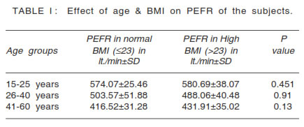
The PEFR values of 300 subjects had been stratified
according to age and BMI. The mean PEFR in
different age groups showed a decreasing trend with
advancement of age. The mean PEFR in age group
of 25-40 years was found to be 577.5+32.49 L/min.
whereas that in the age group of 41-60 years was
426.85+34.5 L/min (Fig. 1). Similarly PEFR appeared
to be higher in normal BMI subjects(mean PEFR
485+73.49 L/min.) than that in the higher BMI
subjects (mean PEFR 474.05+64.41 L/min.). Further
in-depth analysis using Student's t test after
stratification of the high and normal BMI subjects in
various age ranges showed that mean PEFR had
been found to be lower in high BMI subjects in all
age groups except in younger age range of 15-25
years but the differences were not significant
statistically (P value >0.05) as evident in Table I. |
 |
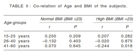
Linear regression analysis model shows that the age significantly affects the PEFR unlike BMI. Age as an independent predictor predicts the 72.3% variability (R2 is 72.3%) in PEFR while BMI > 23 as an independent predictor predicts only 1.4% variability (R2 is 0.014). PEFR was found to be significantly decreased (p value 0.002 & 0.000 respectively) in the age range of 26-40 years and 41-60 years. The odd's ratio for PEFR in different age groups i.e. 1525 years, 26-40 years & 41-60 years are -0.850, -3.207 & -4.082 respectively. This means the decrease in PEFR in age group 41-60 years is more than that in the age group 26-40 years and the decrease in PEFR in the age group 26-40 years is more than in age group 15-25 years. Although with increase in BMI PEFR is decreasing but it is not significant (p=0.855). Table II shows the correlation between the PEFR of
normal and high BMI subjects in various age ranges.
The only significant correlation was found to be in
the age range of 41-60 years (p=0.01) where high
BMI was found to be negatively correlated with PEFR
although the strength of association was poor
(r=–0.244). In other age groups the correlation was
not significant. |
 |
The interplay between age and BMI has been found to be very complex in context of its overall effect on PEFR values. PEFR, on the other hand, is an important diagnostic and prognostic tool in lung function studies for identifying airflow limitations, its severity and variations. Age has been found to be an important factor determining PEFR in healthy subjects. Increasing age appears to be negatively correlated with PEFR although the strength of association is poor. This finding is in agreement with various Indian and foreign authors like Mahajan et al (4), Jepegnanam et al (5), Yogesh Saxena et al (6), Joffa Paul et al (7). The reason for this decline could be related to various factors which plays important role in determining PEFR like expiratory muscle effort, elastic recoil and airway size etc. These factors are known to be affected significantly in old age or as the age advance after 40 years (8). The decline in PEFR per decade increase in age in previous Indian studies ranged between 20.3 to 33.1 L/min (9-13). In a study by Rajendra Prasad et al (14), the decline in PEFR per decade increase in age was 29.2 lit/min. Increase in BMI did not show any significant correlation with mean PEFR in younger age range. These findings show agreement with previous studies (3, 6, 7). In elderly age group i.e. >40 years of age PEFR shows some significant declining trend with higher BMI. besides decrease in the elastic recoil of the lungs and airway size and weakness of the respiratory muscles in older age group, it could further be explained through several possible mechanisms such as mechanical effects on the diaphragm and fat deposition between the muscles and the ribs that can lead to increase in the metabolic demands and work load of breathing (6). Limitations Because of resource limitations, this study has been done as a hospital based study; therefore the study population may not be the true representative of Indian population. There is a scope of population based epidemiological study with bigger sample size to further validate the findings of the study. Conclusion Both age and BMI independently affects the PEFR in Indian Populations but the effect of age on PEFR is much more significant than BMI. Thus, pulmonary functions vary according to the physical characteristics like age and BMI with regional differences in lung functions in healthy Indians. |
|